
FIGURE
Fig. 3
- ID
- ZDB-FIG-110720-59
- Publication
- White et al., 2011 - A transgenic zebrafish model of targeted oocyte ablation and de novo oogenesis
- Other Figures
- All Figure Page
- Back to All Figure Page
Fig. 3
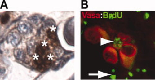
Immunohistochemical detection of BrdU incorporation and germline markers in ovarian tissue, indicating germline cell proliferation, following 14 days Mtz treatment (5 mM). A: Clusters of BrdU-positive cells were readily detectable at end of treatment period. B: Immunohistochemical identification of a candidate oogonial stem cell (arrowhead) positive for both BrdU (green) and the germline marker Vasa (Red); arrows indicate BrdU-positive somatic cells. |
Expression Data
| Gene: | |
|---|---|
| Antibody: | |
| Fish: | |
| Anatomical Term: | |
| Stage: | Adult |
Expression Detail
Antibody Labeling
Phenotype Data
Phenotype Detail
Acknowledgments
This image is the copyrighted work of the attributed author or publisher, and
ZFIN has permission only to display this image to its users.
Additional permissions should be obtained from the applicable author or publisher of the image.
Full text @ Dev. Dyn.