
- Title
-
A transgenic zebrafish model of targeted oocyte ablation and de novo oogenesis
- Authors
- White, Y.A., Woods, D.C., and Wood, A.W.
- Source
- Full text @ Dev. Dyn.
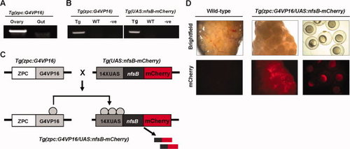
Generation of Tg(zpc:G4VP16/UAS:nfsB-mCherry) transgenic line. A: Ovary-specific G4VP16 mRNA expression was confirmed in adult Tg(zpc:G4VP16) females by RT-PCR. B: Genomic PCR of transgene inserts, zpc:G4VP16 and UAS:nfsB-mCherry, confirming bitransgenic genotype. C: Schematic diagram demonstrating cis-activation of nfsB-mCherry expression by zpc:G4VP16 in Tg(zpc:G4VP16/UAS:nfsB-mCherry) transgenic line. D: nfsB-mCherry expression in Tg(zpc:G4VP16/UAS:nfsB-mCherry) transgenics was confirmed by the detection of mCherry in ovarian tissues (oocytes), and by thepresence of mCherry protein in spawned eggs. |
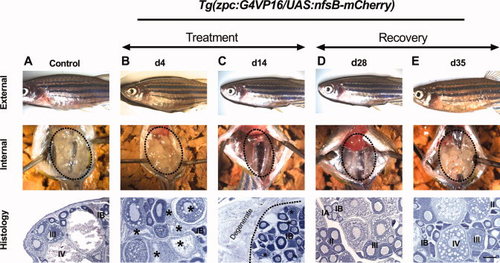
Ovarian degeneration and regeneration following Mtz treatment. A: Representative micrograph demonstrating gross morphology (external, internal) and ovarian histology of untreated (control) fish; note distended abdomen, large ovarian lobes and abundant oocytes at multiple stages of development throughout ovary. B:Tg(zpc:G4VP16/UAS:nfsB-mCherry) female after 4 days exposure to Mtz (5 mM); external appearance and internal ovarian morphology are similar to that of untreated controls; histologically, analysis reveals evidence of degeneration in large oocytes (*). C:Tg(zpc:G4VP16/UAS:nfsB-mCherry) female after 14 days exposure to Mtz (5 mM); note severe ovarian atrophy, exhibited by loss of abdominal distention and recognizable oocytes. Histological analysis revealed extensive ovarian (oocyte) degeneration, and clusters of early-stage oocytes. D:Tg(zpc:G4VP16/UAS:nfsB-mCherry) female 14 days after Mtz removal (experiment d28); ovarian regeneration is observed. E:Tg(zpc:G4VP16/UAS:nfsB-mCherry) female 21 days after Mtz removal (experiment day 35); external appearance, ovarian morphology and histological features are indistinguishable from controls. Oocyte stages are as described in Selman et al. (1993). Degenerate oocytes are indicated by *; dashed line indicates abdominal morphology; ovarian lobes are outlined. Scale bar = 50 μm. |
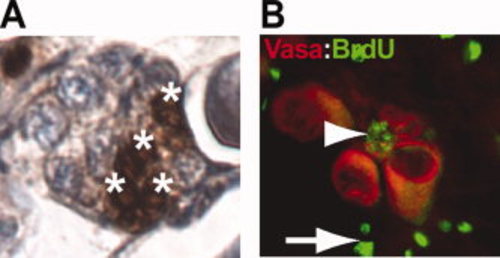
Immunohistochemical detection of BrdU incorporation and germline markers in ovarian tissue, indicating germline cell proliferation, following 14 days Mtz treatment (5 mM). A: Clusters of BrdU-positive cells were readily detectable at end of treatment period. B: Immunohistochemical identification of a candidate oogonial stem cell (arrowhead) positive for both BrdU (green) and the germline marker Vasa (Red); arrows indicate BrdU-positive somatic cells. EXPRESSION / LABELING:
|