
Fig. 4
- ID
- ZDB-FIG-101119-9
- Publication
- Distel et al., 2009 - Optimized Gal4 genetics for permanent gene expression mapping in zebrafish
- Other Figures
- All Figure Page
- Back to All Figure Page
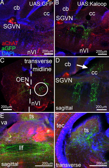
Rh3/5-Kalooping identifies the secondary octaval nucleus (SON) as rhombomere 5-derived. Immunhistochemistry for GFP (green) and Cholinacetyltransferase (ChAT; red) on brain vibratome sections of adult zebrafish derived from crosses of (A) rh3/5:KalTA4 × 4xKGFP and (B–F) rh3/5:KalTA4 × 4xKaloop. All nuclei were counterstained with DAPI (blue). (A and B) Sagittal sections of the hindbrain. (C) Transverse section through the caudal GFP-expressing hindbrain region (r5) in B. (midline marked by dashed line). (D) Sagittal section (rostral is left) showing GFP-positive dendrites in the crista cerebellaris (white arrow). (E) Axons of GFP-positive neurons project through the midbrain into the (F) torus semicircularis. Note typical periventricular cholinergic cells in optic tectum. Abbr.: cb: corpus cerebelli, cc: crista cerebellaris, llf: lateral longitudinal fascicle, nVI: rostral and caudal abducens nuclei, OEN: octavolateralis efferent neurons, r: rhombomere, SGVN: secondary gustatory/viscerosensory nucleus, ts: torus semicircularis, tec: optic tectum, va: valvula cerebelli. |