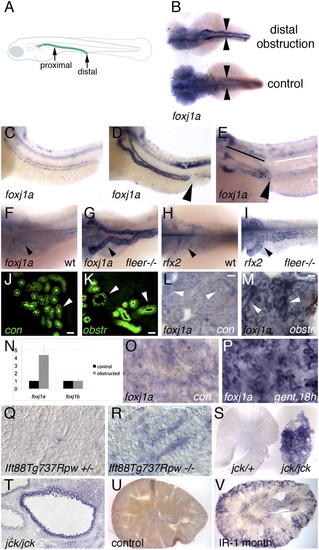
Up-regulation of foxj1a expression in epithelial injury and cystic distension. Embryos in B-I were all imaged at 56 hpf. (A) Schematic diagram illustrating location of the zebrafish pronephric duct (green line) and sites of mechanical proximal and distal obstruction. (B) foxj1a is strongly up-regulated in pronephros of distally obstructed embryos (top), whereas baseline expression is low in paired pronephric ducts (arrowheads) of unobstructed control embryos (bottom). Dorsal views of embryos are shown. Treatment with cycloheximide does not prevent obstruction-mediated increase in foxj1a expression (D) compared with unobstructed embryos (C). (E) Proximal pronephric obstruction (arrowhead) results in up-regulation of foxj1a only in dilated regions proximal to site of obstruction (black line) and not in tubule cells distal to obstruction (white line). (F–I) Both foxj1a and rfx2 are up-regulated in zebrafish cystic kidney mutants. (F) In WT (wt) embryos, foxj1a was expressed at low level in pronephric epithelial cells. (G) foxj1a expression was strongly up-regulated in dilated pronephric tubules of fleer mutant tubules, which develop cysts. Similarly, rfx2 gene expression was up-regulated in dilated kidney tubules of fleer mutants (I) compared with controls (H). (J-M) Obstruction-mediated induction of foxj1a expression in adult zebrafish kidney. Distal mesonephric obstruction in adult zebrafish results in tubule dilation after 12 h (K) compared with control sham-operated kidneys (J) visualized using the Tg(atase1a1a.4:eGFP) transgenic line (30). Whole-mount in situ hybridization of adult zebrafish kidney with foxj1a probe shows low level of expression in sham-operated control (L), whereas distal obstruction (M) induces foxj1a expression in mesonephric tubules after 18 h (arrowheads). (Scale bar: N-Q, 25 μm.) (N) Quantification of foxj1a and foxj1b expression by quantitative RT-PCR in obstructed embryos. (O) foxj1a is expressed at low level in whole-mount adult zebrafish kidney, whereas foxj1a is up-regulated in tubule cells after 18 h of treatment with the nephrotoxin gentamicin (P). (Q) Sections of WT post-embryonic day-7 mouse kidneys stained for Foxj1 mRNA expression show low level of Foxj1 RNA, whereas similar sections of Ift88Tg737Rpw-/- mutant mouse kidneys (R) show strong cortical expression of Foxj1 in subset of tubule cells. (S) jck/+ Kidney slice (left) compared with slice of cystic jck/jck kidney (right), revealed strong up-regulation of Foxj1 in jck/jck cystic kidney. (T) Section of jck/jck kidney showing strong expression in cyst lining epithelial cells. Increased Foxj1 expression was also observed in mice subjected to renal ischemia-reperfusion injury 1 mo postrecovery (V) compared with control kidneys (U).
|
