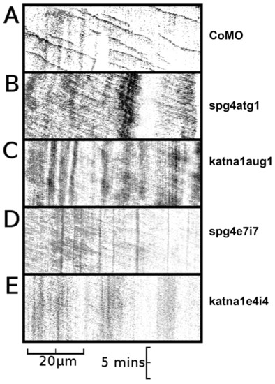
Axons in spast and katna1 morphants lack discrete EB3-GFP puncta. (A) Kymograph of an axon made from a 10-minute time-lapse recording of a neuron from a CoMO-injected embryo, demonstrating anterograde movement of discrete comet-like puncta of EB3-GFP fluorescence (CoMO, n=21; spg4CoMO, n=12). (B) Kymograph of an axon made from a 10-minute time-lapse recording of a neuron from a spg4atg1 (0.6 pmol)-injected embryo, demonstrating weak anterograde movement of diffuse GFP fluorescence in the absence of discrete comet-like puncta of EB3-GFP fluorescence (n=13). (C) Kymograph of an axon made from a 10-minute time-lapse recording of a neuron from a katna1aug1 (1.8 pmol)-injected embryo, again demonstrating weak anterograde movement of diffuse GFP fluorescence in the absence of discrete puncta of EB3-GFP fluorescence (n=10). Equivalent results were obtained with the spg4e7i7 [1.2 pmol; n=6; (D)] and katna1e4i4 [1.2 pmol; n=6; (E)] morpholinos. Horizontal scale bar: 20 µm, vertical scale bar: 5 minutes. A series of individual frames from each of these time-lapse recordings is shown in supplementary material Fig. S4. See supplementary material Movies 2-6.
|
