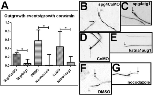
Disruption of microtubule dynamics reduces growth cone activity in the zebrafish embryonic CNS. An outgrowth event was scored as a continuous period of growth of a single process from the distal end of an axon. Scoring was carried out on 10-minute time-lapse recordings and the mean number of outgrowth events per growth cone was calculated. (A) The number of outgrowth events occurring per growth cone per minute is reduced by knockdown of spast (P=0.0179), knockdown of katna1 (P=0.025) and treatment with nocodazole (P=0.0155). Values were calculated using a two-tailed Student’s t-test assuming unequal variance. (B–G) Frames taken from 10-minute time-lapse recordings of the distal part of EB3-GFP-expressing neurons in live zebrafish embryos. When embryos were injected with spastin control morpholino (B), standard control morpholino (D), or treated with DMSO (F), growth cones were highly motile and rapid outgrowth and branching of GFP-labelled processes (arrows in B and F) was observed. When embryos were injected with spast (C) or katna1 (E) morpholinos, or treated with nocodazole (G), growth cone activity was greatly reduced. In some cases, the tips of immobile processes (arrows in C and G) were brightly labelled with an accumulation of EB3-GFP. Scale bars: 10 μm. See supplementary material Movies 9-14.
|
