
Fig. 2
- ID
- ZDB-FIG-101105-31
- Publication
- Chitramuthu et al., 2010 - Molecular cloning and embryonic expression of zebrafish PCSK5 co-orthologues: Functional assessment during lateral line development
- Other Figures
- All Figure Page
- Back to All Figure Page
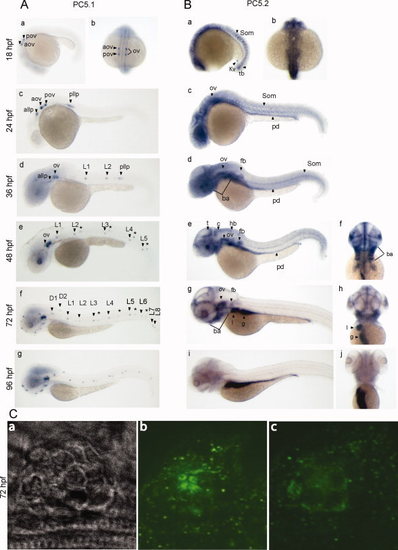
Developmental expression of zebrafish PC5.1 and PC5.2. A: An ontogeny of expression for PC5.1 by whole mount in situ hybridization. Lateral (a) and dorsal (b) view of 18-hr post-fertilization (hpf) embryos revealed very discrete expression within the anterior and posterior part of the otic vesicle. At 24hpf (c), in addition to the anterior and posterior part of the otic vesicle, expression is observed within the anterior and posterior lateral line primordia. Lateral view of 36hpf (d) detected additional expression within the posterior lateral line neuromast L1 and L2. At 48hpf (e), 72hpf (f), and 96 hpf (g), PC5.1 is strongly localized in the otic vesicle and within the anterior and posterior lateral line neuromasts. The asterisks (A: e, f) represent neuromasts on the opposite side of the embryo. B: An ontogeny of expression for PC5.2 by whole mount in situ hybridization. At 18hpf (a, lateral; b, dorsal), ubiquitous expression with distinct regionalization within the somites, Kupffer′s vesicle, and in the tail bud is observed. Lateral view of 24hpf (c) shows continued expression within the somites and CNS with additional expression within the otic vesicle and pronephric duct. At 36hpf (d) and 48hpf (e, lateral view; f, dorsal view), PC5.2 expression is found within the pharyngeal region, fin bud, CNS, otic vesicle, and pronephric duct. Lateral (g, i) and dorsal (h, j) views of 72hpf and 96hpf, respectively, show a higher level of expression within the liver and gut. C: Localization of immunoreactive PC5.1 within the hair cells and supporting cells of the neuromast. Zebrafish embryos at 72hpf were stained with anti-PC5.1 antibody. Bright field image of neuromast (a), localization of PC5.1 within the hair cells (b), and localization of PC5.1 within the supporting cells (c) were visualized using confocal microscopy. A: Aov, anterior part of the otic vesicle; pov, posterior part of the otic vesicle; allp, anterior lateral line primordium; pllp, posterior lateral line primordium; alln, anterior lateral line neuromast; D1 and D2, dorsal lateral line neuromasts; L1-L8, posterior lateral line neuromasts. B: som, somites; Kv, kupffer′s vesicle; tb, tail bud; ov, otic vesicle; pd, pronephric duct; ba, branchial arches; fb, fin bud; c, cerebellum; t, tectum; hb, hindbrain. |