
Fig. 7
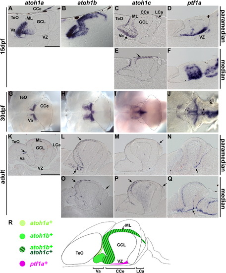
Expression of proneural genes in juvenile and adult cerebellum. Expression of atoh1a, atoh1b, atoh1c, and ptf1a in cerebellum at late larval (15 dpf, A–F), juvenile (30 dpf, G–J), and adult (90 dpf or older, K–Q) stages by in situ hybridization. Sagittal sections, at paramedian (A–D, K–N) or median (E, F, O–Q) levels. Whole-mount in situ hybridization with dorsal views (G–J). Note that the expression domains of atoh1b and atoh1c in the midline are very thin, and were not detected by whole mount in situ hybridization (H, I). (R) Schematic representation of atoh1a, atoh1b, atoh1c, and ptf1a expression in the adult cerebellum. The abbreviations are defined in [Fig. 1] and [Fig. 3]. Scale bars: 100 μm (A), 500 μm (G), 1 mm (K). |
| Genes: | |
|---|---|
| Fish: | |
| Anatomical Terms: | |
| Stage Range: | Days 14-20 to Adult |
Reprinted from Developmental Biology, 343(1-2), Kani, S., Bae, Y.K., Shimizu, T., Tanabe, K., Satou, C., Parsons, M.J., Scott, E., Higashijima, S.I., and Hibi, M., Proneural gene-linked neurogenesis in zebrafish cerebellum, 1-17, Copyright (2010) with permission from Elsevier. Full text @ Dev. Biol.