ZFIN ID: ZDB-FIG-100525-7 |
Gene: | |
---|---|
Fish: | |
Anatomical Terms: | |
Stage: | Pec-fin |
Fish: | |
---|---|
Observed In: | |
Stage: | Pec-fin |
Fig. 6 
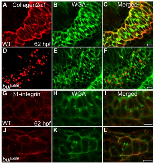
Sec24D-deficient chondrocytes do not transport ECM proteins. (A–F) Collagen2α1 (red) and Wheat Germ Agglutinin (WGA, green) signals are concentrated at juxtanuclear compartments and in the extracellular space of wild-type chondrocytes (A–C). In bulldog, the signals are primarily localized in cytoplasmic vesicular compartments, although weak WGA signal is also detectable at the plasma membrane and the extracellular space (D–F). (G–L) Single pass confocal images of wild-type and bulm606 Meckel′s cartilages labeled with the β1-integrin (red) recognizing antibody and counterstained with WGA (green). Merged channels show co-localization of the two labels in the cell boundaries in both wild-type and bulldog chondrocytes. The right panels represent merged images of the left and middle panels. Scale bars are 5 μM. |
Gene | Antibody | Fish | Conditions | Stage | Anatomy | Assay |
---|---|---|---|---|---|---|
col2a1a | AB | standard conditions | Pec-fin | pharyngeal arch cartilage extracellular matrix | IHC | |
sec24dm606/m606 (AB) | standard conditions | Pec-fin | chondrocyte cytoplasm | IHC |
Fish | Conditions | Stage | Phenotype |
---|---|---|---|
sec24dm606/m606 (AB) | standard conditions | Pec-fin | extracellular matrix constituent secretion decreased occurrence, abnormal |
Acknowledgments: | This image is the copyrighted work of the attributed author or publisher, and ZFIN has permission only to display this image to its users. Additional permissions should be obtained from the applicable author or publisher of the image. Full text @ PLoS One |
---|