
Fig. 1
- ID
- ZDB-FIG-100422-54
- Publication
- Schonthaler et al., 2010 - The zebrafish mutant bumper shows a hyperproliferation of lens epithelial cells and fibre cell degeneration leading to functional blindness
- Other Figures
- All Figure Page
- Back to All Figure Page
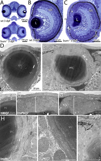
The lens in 3 dpf bum-/- zebrafish shows the onset of a hyperproliferation of the anterior epithelium. (A–C) Toluidine blue-stained transversal semi-thin sections through the central part of the eyes of 3 dpf sibling (wild-type, wt; top picture in A and B) and bum-/- mutant zebrafish larvae (bottom picture in A and C). (A) Cross-sections of entire heads, (B and C) higher magnifications of individual eyes. Note the onset of a hyperproliferation of the anterior lens epithelium in the mutant (C; white arrowheads). The overall morphology of the eye however, including the layers of the neural retina (NR), is normal in bum-/- mutant larvae (compare B and C). (D–J) Thin-section electron microscopy images of transversal sections through the central part of the eyes of 3 dpf sibling (D, F) and bum-/- (E, G–J) zebrafish larvae. (D, E) Overviews showing the morphology of the entire lens. (F, G) Higher magnifications of the anterior lens epithelia in sibling (F) and bum-/- (G, G′) zebrafish larvae. Note that the epithelium (brackets) forms a monolayer in the wild-type larvae, but is expanded to several cell layers in the mutants. Occasionally, apoptotic cells can be observed in the mutant lens epithelium (asterisk in G′). (H, I) Higher magnifications of the lens’ bow region in bum-/- mutant zebrafish larvae showing the differentiating secondary lens fibre cells, which at this stage do not yet show the massive degenerative changes observed at later developmental stages (see [Fig. 2] and [Fig. 4]). (J) Slight deformation of the outer cortex of a bum-/- mutant lens anterior of the lens’ equator. Asterisks denote superficial secondary lens fibre cells that show the first signs of cellular swelling. Abbreviations: C, cornea; PFC, primary lens fibre cells; L, lens; LE, lens epithelium; n, secondary lens fibre cell nuclei; NR, neural retina; ON, optic nerve; P, pigment granules of the iris; PFC, primary lens fibres; SFC, secondary lens fibre cells. |
| Fish: | |
|---|---|
| Observed In: | |
| Stage: | Protruding-mouth |
Reprinted from Mechanisms of Development, 127(3-4), Schonthaler, H.B., Franz-Odendaal, T.A., Hodel, C., Gehring, I., Geisler, R., Schwarz, H., Neuhauss, S.C., and Dahm, R., The zebrafish mutant bumper shows a hyperproliferation of lens epithelial cells and fibre cell degeneration leading to functional blindness, 203-219, Copyright (2010) with permission from Elsevier. Full text @ Mech. Dev.