|
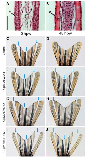
Interray wound-healing assay demonstrating the role of IGF and Activin/TGFβ signaling in wound closure. (A,B) Hematoxylin and Eosin staining of longitudinal fin sections. (A) Interrays lack skeletal elements, have multilayered epidermis, and very loose mesenchyme. (B) Rays contain bones covered by thin epidermis. Mesenchyme appears as a compact aggregation of interconnected fibroblasts. (C-J) Bright-field images of injured fins. Arrows indicate incisions between the bony rays. These interray notches were closed after 48 hours in control fins (C,D). Exposure to either NVP-AEW541 (E,F) or NVP-ADW742 (G,H) markedly impaired wound healing. Treatment with the inhibitor of Activin/TGFβ signaling, SB431542, completely blocked wound closure (I,J). b, bones; e, epidermis; m, mesenchyme; hpw, hours post-wounding. Scale bar: 50 μm.
|