
Fig. S4
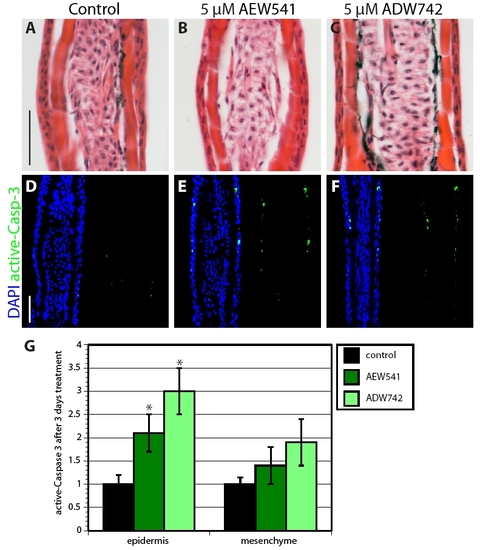
Inhibition of IGF signaling increases cell apoptosis in uninjured epidermis. (A-C) Hematoxylin-Eosin stained rays of uninjured fins that were treated with 0.05% DMSO (A), NVP-AEW541 (B) or NVP-ADW742 (C) do not display differences in morphology of the epidermis. (D-F) Sections of uninjured fins stained with active-Caspase-3 antibody in green and with the nuclear marker DAPI in blue. IGF-1R inhibitor-treated fins (E,F) contain a few more active-Caspase-3-positive cells in the epidermis than in the control (D). (G) Quantification of active-Caspase-3 in uninjured fins treated with IGF-1R inhibitors for 3 days. n=6; P<0.01. Scale bars: 50 μm. |