
Fig. S3
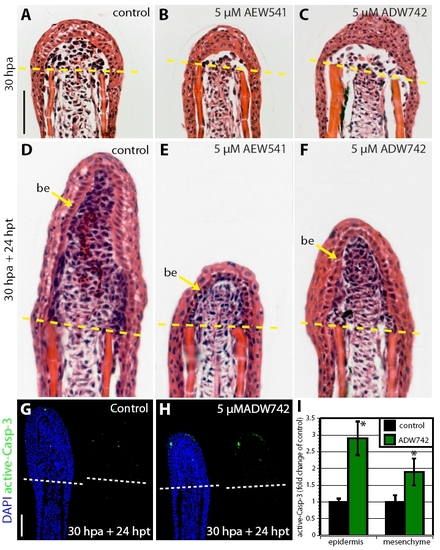
The effect of IGF-1R signaling on the wound epidermis. (A-F) Hematoxylin-Eosin stained fin sections. (A-C) At 30 hpa, the basal layer of the wound epidermis of the control fin (A) appears more ordered and aligned than in fin treated with NVP-AEW541 (B) or NVP-ADW742 (C). (D-F) Drug-shift experiment: 30 hours at normal conditions and 24 hours with treatment. The architecture of the wound epidermis appears similar in control (D) and inhibitor-treated (E,F) fins. (G,H) Drug-shift experiment (30 hours at normal conditions and 24 hours with treatment). Sections of regenerates stained with active-Caspase-3 antibody in green and a nuclear marker DAPI in blue. Control fins (G) and ADW742-treated fins (H) contain low amounts of active-Caspase-3-positive cells. (I) Quantification of active-Caspase-3 in control and inhibitor-treated fins in the drug-shift experiment. n=6; P<0.01. Scale bars: 50 μm. |