|
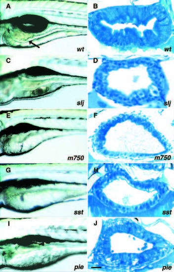
Mutations affecting the developing zebrafish intestine. (A,C,E,G,I) Lateral views of living 5 dpf larvae. (B,D,F,H,J) Corresponding histological cross-sections through the mid-intestine. Lumen size varies with position along the anterior-posterior axis and with muscular contractions. (A,B) Wild type. The intestinal epithelium (arrow) is highly folded, bile is visible in the lumen and the gas bladder is inflated. The intestinal epithelium is polarized with basal nuclei and a microvillus brush border. (C,D) slj m74, (E,F) m750, (G,H) sstm311, (I,J) piem497. The intestinal wall of all mutants is thin, lacks folds and the epithelium is not fully polarized and shows signs of degeneration. Scale bar, B, F and H, 20 μm; D, 15 μm; J, 25 μm.
|