|
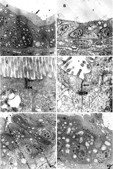
Electron micrographs showing regional intestinal differentiation. (A,C,E) wild type and (B,D,F) sljm74. (A) The wild-type anterior intestinal epithelium is columnar and has a prominent microvillus brush border (arrow). (B) sljm74 anterior intestinal epithelial cells are cuboidal with few microvilli. (C,D) Adherens (arrowhead) and tight junctions (arrow) are present in the anterior intestine of both (C) wild type and (D) sljm74. (E,F) Posterior intestinal epithelium appears normal in both (E) wild type and (F) sljm74. * labels the intestinal lumen. Magnification: A, x950; B, x1100; C, x22,000; D, x28,500; E, x2200; F, x2950.
|