|
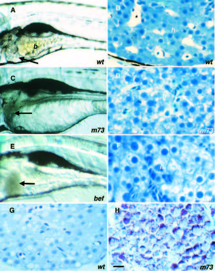
Liver mutants. (A,BG) Wild type. (C,D,H) m73. (E,F) bef m362. (A) Lateral view of wild-type 5 dpf larva with bile (b) in intestinal lumen; arrow points to liver. (B) Cross section of wild-type liver 5 dpf with erythrocytes in the sinusoids (*) and hepatocytes (h). (C) The day-4 m73 liver (arrow) has a gray-brown hue. (D) m73 hepatocytes are degenerating and erythrocytes (arrow) are pooling in the sinusoids. (E) befm362 liver (arrow) has a red-brown hue. (F) befm362 hepatocytes also degenerate and compress the sinusoids. (G) Wild-type liver 6 dpf does not stain with PAS. (H) m73 liver 6 dpf stains for PAS. Scale bar, 15 μm.
|