|
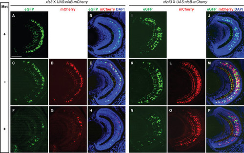
Prodrug dependent ablation of specific bipolar cell types in larvae from enhancer trap lines. (A-H) Confocal images of retinal cross-sections from larvae (5 dpf) produced by mating of UAS:nfsB-mCherry transgenic fish to Tg(Gal4-VP16;UAS:eGFP)xfz3. (A, B) Retina from a larva without the UAS:nfsB-mCherry fusion gene that was unaffected by Met treatment. (C-E) Retina from larva expressing NTR-mCherry (red) in bipolar cells. (F-H) Retina from larva expressing NTR-mCherry (red) showed loss of fluorescently labelled bipolar cells following Met treatment. (I-P) The same type of analysis as in (A-H) conducted on larvae produced by mating of UAS:nfsB-mCherry transgenic fish to Tg(Gal4-VP16;UAS:eGFP)xfz43. (N-P) Significant degeneration and loss of labelled bipolar cells occurred in the retina of Met treated larvae. Labelling to the left indicate Met treatment (+) and no treatment (-). The crosses and different types of fluorescence are indicated at the top. Scale bar: 50 μm.
|