
Fig. 5
- ID
- ZDB-FIG-090515-31
- Publication
- Riley et al., 1996 - A mutation in zebrafish affecting a localized cellular function required for normal ear development
- Other Figures
- All Figure Page
- Back to All Figure Page
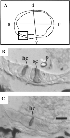
Sectional analysis of monolith. Sections of utricular sensory epithelia in histochemically stained 4-day chimeras. A number of chimeras initially analyzed by fluorescence imaging (Fig. 3) were fixed at 4 days, histochemically stained to visualize labeled +/+ cells, and sectioned. A total of 48 sectioned ears were analyzed. (A) Location of the utricular sensory epithelium within the ear. (B) The utricular sensory epithelium of a normal ear in which one hair cell and one support are labeled. The posterior edge of the utricular otolith is indicated. Of 21 normal ears that were sectioned, 15 contained labeled cells in the anteroventral epithelium; all 15 contained both labeled hair cells and labeled support cells in this region. For these ears, the average number (mean ± SD) of labeled cells visible in this region was 1.6 ± 0.7 hair cells, 1.3 ± 0.4 support cells, and 3.0 ± 1.1 total cells. (C) The utricular sensory epithelium of an abnormal ear in which one hair cell is labeled. A total of 8 abnormal ears with anteroventral fluorescence were sectioned; all contained histochemically stained hair cells in this region, but none contained labeled support cells. For these ears, the average number of labeled cells visible in this region was 2.0 ± 0.9 hair cells, 0 ± 0 support cells, and 2.2 ± 1.3 total cells. Nineteen abnormal ears in which no fluorescently labeled +/+ cells were detected in the anteroventral quadrant at 48 hr were also sectioned. Consistent with the early analysis, no histochemically stained +/+ cells were evident in any of these sections (not shown). Abbreviations: hc, hair cell; o, otolith; sc, support cell. In all panels, anterior is toward the left and dorsal is toward the top. The scale bar corresponds to 10 μm for B and C. |
Reprinted from Developmental Biology, 179(2), Riley, B.B. and Grunwald, D.J., A mutation in zebrafish affecting a localized cellular function required for normal ear development, 427-435, Copyright (1996) with permission from Elsevier. Full text @ Dev. Biol.