
Fig. 1
- ID
- ZDB-FIG-090515-28
- Publication
- Riley et al., 1996 - A mutation in zebrafish affecting a localized cellular function required for normal ear development
- Other Figures
- All Figure Page
- Back to All Figure Page
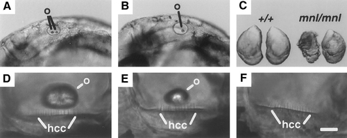
Morphological analysis of monolith. (A) A 1-day (day of development) +/+ embryo showing two otoliths (o) in the inner ear. (B) A 1-day mnl/mnl embryo with only one otolith in the inner ear. (C) Otoliths from similarly sized +/+ and mnl/mnl adults. Ventral surfaces that contact the sensory epithelia are shown. (D) Utriculus of a 4-day +/+ embryo with a normal otolith (o). (E) Utriculus of a 4-day mnl/mnl embryo with an abnormally small otolith (o). (F) Utriculus of a 4-day mnl/mnl embryo with no otolith. Hair cell cilia (hcc) are evident in the sensory epithelia of each utriculus. In A, B, and D–F, anterior is toward the left and dorsal is toward the top. The scale bar corresponds to 120 μm for A and B, 570 μm for C, and 15 μm for D–F. |
Reprinted from Developmental Biology, 179(2), Riley, B.B. and Grunwald, D.J., A mutation in zebrafish affecting a localized cellular function required for normal ear development, 427-435, Copyright (1996) with permission from Elsevier. Full text @ Dev. Biol.