
Fig. 3
- ID
- ZDB-FIG-090515-30
- Publication
- Riley et al., 1996 - A mutation in zebrafish affecting a localized cellular function required for normal ear development
- Other Figures
- All Figure Page
- Back to All Figure Page
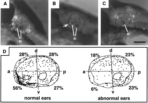
Chimeric analysis of monolith. One hundred ninety-eight +/+ → mnl/mnl chimeras were screened at 24 hr for otolith morphology (Table 1) and further analyzed by fluorescence imaging at 48 hr. (A) A normal ear with numerous labeled cells throughout the inner ear. The locations of cells that potentially facilitated phenotypic rescue could not be inferred from such widespread colonization patterns. (B) A normal ear in which a single labeled cell (or small group of cells) is visible in the anteroventral quadrant of the ear (arrow). (C) An abnormal ear with several labeled cells in the posterior half of the ear. The label visible near the anterodorsal border of the ear marks a neuron in the hindbrain. (D) Frequency and distribution of labeled cells in chimeric ears. The percentage of ears with labeled cells in the indicated quadrants is shown. After excluding ears with ubiquitous labeling, such as that shown in A, positions of labeled cells (dark spots) were projected onto maps representing 89 normal and 79 abnormal ears, respectively. The scale bar corresponds to 100 μm for A– C and 40 μm for D. |
Reprinted from Developmental Biology, 179(2), Riley, B.B. and Grunwald, D.J., A mutation in zebrafish affecting a localized cellular function required for normal ear development, 427-435, Copyright (1996) with permission from Elsevier. Full text @ Dev. Biol.