
Fig. 2
- ID
- ZDB-FIG-090515-29
- Publication
- Riley et al., 1996 - A mutation in zebrafish affecting a localized cellular function required for normal ear development
- Other Figures
- All Figure Page
- Back to All Figure Page
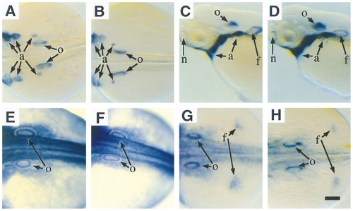
Molecular analysis of monolith. (A–D) Whole mount in situ hybridizations of 24 hr +/+ (A), 24 hr mnl/mnl (B), 48 hr +/+ (C), and 48 hr mnl/mnl embryos (D) with riboprobe specific for dlx3. (E, F) Whole mount in situ hybridizations of 24 hr +/+ (E) and 24 hr mnl/mnl embryos (F) with riboprobe specific for msxC. (G, H) Whole mount in situ hybridizations of 24 hr +/+ (G) and 24 h mnl/mnl embryos (H) with riboprobe specific for msxD. All panels show dorsal views except C and D, which show lateral views. Anterior is toward the left in all panels. Abbreviations: a, pharyngeal arches; f, fin buds; n, nasal pits; o, otic vesicles. The scale bar corresponds to 100 μm for A, B, and E–H and 150 μm for C and D. |
Reprinted from Developmental Biology, 179(2), Riley, B.B. and Grunwald, D.J., A mutation in zebrafish affecting a localized cellular function required for normal ear development, 427-435, Copyright (1996) with permission from Elsevier. Full text @ Dev. Biol.