|
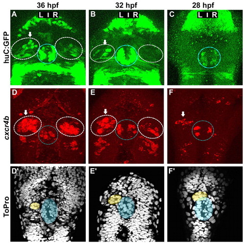
Asymmetric cxcr4b expression precedes the migration of the parapineal and the appearance of HuC+ habenular neurons. (A-C) Confocal projections of GFP fluorescence in Tg(huC:gfp) zebrafish embryos at 36 (A), 32 (B) and 28 (C) hpf. (D-F′) Confocal projections of fluorescent in situ hybridisations showing cxcr4b transcripts (D-F) and ToPro nuclear staining in the corresponding embryos (D′-F′) at 36 (D,D′), 32 (E,E′) or 28 (F,F′) hpf. Nuclear staining permits detection of the parapineal (yellow) and pineal (blue). In A-F, the habenulae are indicated by a dashed white line, the epiphysis by a blue line. Arrows indicate the habenula with the higher number of huC:GFP-positive habenular neurons or cxcr4b-expressing cells.
|