
Fig. 2
- ID
- ZDB-FIG-081219-36
- Publication
- Berndt et al., 2008 - Rho-kinase and myosin II affect dynamic neural crest cell behaviors during epithelial to mesenchymal transition in vivo
- Other Figures
- All Figure Page
- Back to All Figure Page
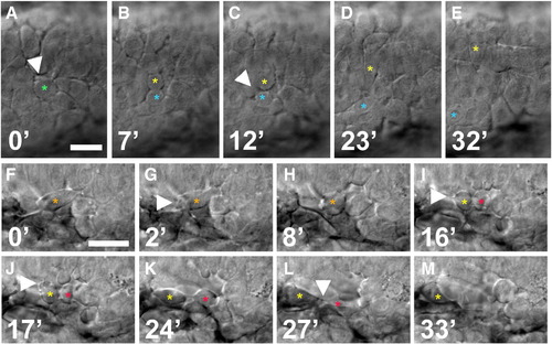
Imaging of cytokinesis in premigratory NCCs. (A–M) Time-lapse sequences in wild-type embryos showing cytokinesis. (A–E) Dorsal views of rhombomere 2 dorsal neuroepithelium. Parent cell is labeled with green asterisk before division and daughters are labeled with blue and yellow asterisks. A parent cell blebs extensively before division (arrowhead in A). Arrowhead in C indicates a filopodium extended from daughter cell. (F–M) Lateral views of cytokinesis in a mesenchymal NCC adjacent to rhombomere 3. Parent cell is labeled with orange asterisk and daughters with red and yellow asterisks. Arrowhead in G indicates blebbing during cell rounding. Arrowhead in I indicates filopodial protrusion prior to the completion of cytokinesis. Arrowhead in J indicates blebbing after cytokinesis. Arrowhead in L indicates a narrow intercellular bridge connecting daughter cells. Time is in minutes. Scale bars = 20 μm. |
Reprinted from Developmental Biology, 324(2), Berndt, J.D., Clay, M.R., Langenberg, T., and Halloran, M.C., Rho-kinase and myosin II affect dynamic neural crest cell behaviors during epithelial to mesenchymal transition in vivo, 236-244, Copyright (2008) with permission from Elsevier. Full text @ Dev. Biol.