
Fig. 4
- ID
- ZDB-FIG-081219-38
- Publication
- Berndt et al., 2008 - Rho-kinase and myosin II affect dynamic neural crest cell behaviors during epithelial to mesenchymal transition in vivo
- Other Figures
- All Figure Page
- Back to All Figure Page
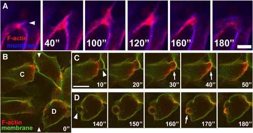
Actin dynamics in protrusions of neuroepithelial cells undergoing EMT. (A) Time-lapse sequence showing that F-actin localization coincides with membrane protrusion during filopodial extension (arrowhead). Cells are expressing GPI-GFP to label membranes (blue) and RFP-UtrCH to label F-actin (red). Dorsal views of basal edge of rhombomere 5/6, anterior is left, embryo is approximately 16 hpf. (B–D) Cells labeled with GPI-GFP (green) and RFP-UtrCH (red) showing actin dynamics in blebs during EMT. B shows location of cells shown in time-lapse in C and D. Arrowheads in B delineate approximate location of edge of neuroepithelium. (C) Cell within the neuroepithelium blebbing at the basal surface. (D) Cell delaminating from the neuroepithelium. During extension, blebs do not contain actin (arrowheads in C, D). During bleb retraction, actin accumulates beneath the membrane (arrows in C, D). Dorsal view of basal edge of rhombomere 4, anterior is up, embryo is approximately 14 hpf. Time is in seconds. Scale bars = 5 μm for A and 10 μm for B–D. |
Reprinted from Developmental Biology, 324(2), Berndt, J.D., Clay, M.R., Langenberg, T., and Halloran, M.C., Rho-kinase and myosin II affect dynamic neural crest cell behaviors during epithelial to mesenchymal transition in vivo, 236-244, Copyright (2008) with permission from Elsevier. Full text @ Dev. Biol.