
Fig. S9
- ID
- ZDB-FIG-081117-16
- Publication
- Aman et al., 2008 - Wnt/beta-catenin and Fgf signaling control collective cell migration by restricting chemokine receptor expression
- Other Figures
- All Figure Page
- Back to All Figure Page
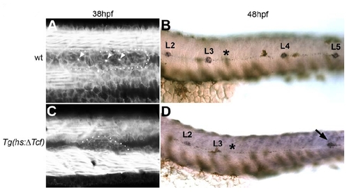
Rosette Formation Is Lost in Migrating Tg(hsΔTCF:GFP) Primordia (A) Rosettes can be visualized in 38hpf wt embryos by focal accumulation of Factin revealed by phalloidin staining (arrowheads). (B) eya1 in situ shows that wt primordia have completed migration and deposited all posterior neuromasts by 48hpf (L2-L4). (C) Heatshock induction of the dominant repressor ΔTCF:GFP at 28hpf leads to complete loss of rosettes by 38hpf as evidenced by phalloidin staining. At 28hpf the primordium is located just posterior to L3 (asterisk). (D) eya1 in situ at 48hpf shows that heatshocked Tg(hsΔTCF:GFP) primordia continue to migrate following the loss of rosettes. Activation of ΔTCF:GFP is more detrimental to the general health of the embryos than activation of dkk. Therefore, heatshocked Tg(hsΔTCF:GFP) primordia stall at around the position of L5 (arrow) and the embryos eventually die. |
Reprinted from Developmental Cell, 15(5), Aman, A., and Piotrowski, T., Wnt/beta-catenin and Fgf signaling control collective cell migration by restricting chemokine receptor expression, 749-761, Copyright (2008) with permission from Elsevier. Full text @ Dev. Cell