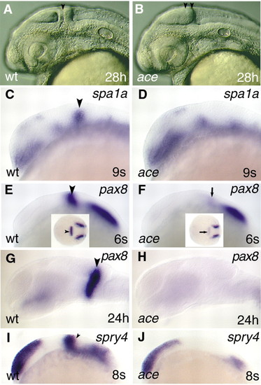
The molecular identity of the isthmic domain is not maintained in ace. All views are rostral to the left, and are lateral apsects, apart from the insets in E and F, which are dorsal views. (A,B) Captures of living embryos illustrating the special anatomical features (lack of isthmus and separate cerebellar anlage) of the ace mutants. The black arrowhead in panel A points to the isthmic constriction; the two black arrowheads in panel B mark the caudal tectal expansion. (C,D) Analysis of spa1a expression reveals the lack of the isthmic expression domain in the mutant embryos in comparison with wild type. The black arrowhead labels the MHB expressing spa1a in wild-type embryo. (E,F) Only a low level of pax8 expression can be detected at the MHB in the mutant embryos (arrows; F,F inset) in comparison with wild types (arrowheads; E,E inset). (G,H) Later on, expression of pax8 is abolished from the prospective MHB region in ace mutants. (I,J) In contrast to pax8, expression of spry4 is not initiated in the mutants. The arrowhead (I) marks the MHB expression domain of spry4 in the wild-type embryo.
|
