FIGURE

Fig. S6

ID
ZDB-FIG-081028-41
Publication
Imamura et al., 2008 - A non-canonical function of zebrafish telomerase reverse transcriptase is required for developmental hematopoiesis
Other Figures
All Figure Page
Back to All Figure Page
Fig. S6

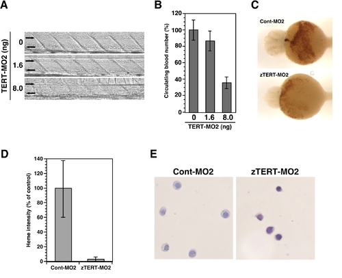
Blood cell number and heme intensity in zTERT-MO2-injected embryos.
(A) Lateral views of the trunk regions of 72 hpf embryos following injection of 0, 1.6, and 8.0 ng of zTERT-MO2. In these bright field pictures, blood cells in the trunks were observed upon a decrease in the injected MO concentrations. The upper vessel is the dorsal artery (left to right arrows) and the lower vessel is the posterior cardinal vein (right to left arrows).
(B) Quantitation of the circulating blood cell number in zTERT-MO2-injected embryos. The percentage of the circulating blood cell numbers at 72 hpf was calculated using uninjected samples (0 ng) as a control and were determined for 15 embryos from each group.
(C) Whole-mount o-dianisidine staining for the presence of heme in Cont-MO2- and zTERT-MO2-injected embryos at 48 hpf (8 ng MOs). Blood flow over the yolk sac and in the tail vessels results in brown staining in Cont-MO2-injected embryos at 48 hpf (ventral view; upper panel), but there is an obvious decrease in this staining following the MO2 injection (lower panel).
(D) Quantification of the heme staining intensity in embryos injected with Cont-MO2 or zTERT-MO2 (8 ng) at 48 hpf. Percentages of the control heme intensity in injected embryos.
(E) Wright-Giemsa staining of isolated blood cells from Cont-MO2- and TERT-MO2-injected embryos at 48 hpf (8 ng MOs).

Expression Data

Expression Detail
Antibody Labeling
Phenotype Data

Phenotype Detail
Acknowledgments
This image is the copyrighted work of the attributed author or publisher, and ZFIN has permission only to display this image to its users. Additional permissions should be obtained from the applicable author or publisher of the image. Full text @ PLoS One