
Fig. 7
- ID
- ZDB-FIG-081028-38
- Publication
- Imamura et al., 2008 - A non-canonical function of zebrafish telomerase reverse transcriptase is required for developmental hematopoiesis
- Other Figures
- All Figure Page
- Back to All Figure Page
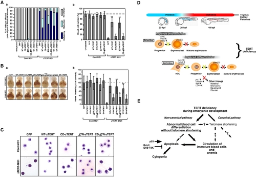
Restoration of ineffective hematopoiesis in TERT-deficient embryos by the expression of zebrafish and human TERT. (A) The blood cell number is rescued in zTERT-MO1-injected embryos (8 ng MOs) at 72 hpf following the injection of the indicated TERT constructs. The percentage of the embryos affected was estimated and assigned into the three categories: No-change, Reduction, or Deficiency, as described above (a). Percentages of control circulating blood cell number in embryos at 72 hpf after the co-injection of several TERT constructs and either Cont-MO1 or zTERT-MO1 at 72 hpf. **P<0.001 (Student t-test) (b). Blood cell numbers were counted in 10 embryos for each group. (B) Rescue of heme appearance, visualized by whole-mount o-dianisidine staining for hemoglobin detection, in TERT-deficient embryos (8 ng MOs) following the injection of several TERT constructs (0.3 ng). Representative samples of whole-mount o-dianisidine staining (a). Percentages of the control heme intensity in embryos at 48 hpf after injection of several TERT constructs with Cont-MO1 or zTERT-MO1 at 48 hpf. *P<0.01, **P<0.001 (Student t-test) (b). Blood cell numbers were counted in 10 embryos for each group. (C) Wright-Giemsa staining of isolated blood cells from Cont-MO1- and TERT-MO1-injected embryos (8 ng) harboring the indicated plasmid constructs (0.3 ng) at 48 hpf. (D) Schematic model of hematopoiesis in TERT-deficient zebrafish embryos. In zebrafish embryos, hematopoiesis occurs in primitive and definitive waves. The first primitive/embryonic wave mainly generates primitive erythrocytes from progenitor cells in the intermediate cell mass (ICM) (represented by the blue color in the 20 and 28 hpf embryo models). The second definitive/adult wave gives rise to hematopoietic stem cells which have the potential to differentiate into all hematopoietic lineages and possess the self-renewal ability to maintain their blood system throughout life. Definitive hematopoietic cells, including stem cells, first arise in the aorta gonad mesonephros (AGM) region (represented by the red color in the 48 hpf embryo model). Definitive hematopoietic stem cells are thought to subsequently colonize the kidney, thymus, and pancreas [53], [88]. Our current model suggests that zTERT deficiency affects both primitive and definitive hematopoiesis in zebrafish, and induces impaired differentiation of the blood cells, including the erythrocyte lineage, prior to maturation. An ‘X’ next to the indicated gene denotes downregulated expression. The yellow ‘X’ in this schematic indicates a somewhat impaired pathway and the red ‘X’ designates a severely impaired pathway. (E) A TERT deficiency in the zebrafish embryo leads to abnormal differentiation and apoptosis, presumably of hematopoietic stem or progenitor cells. This subsequently leads to the circulation of immature blood cells with hypochromic anemia due to a disruption of both primitive and definitive hematopoiesis without telomere shortening. Non-canonical functions of TERT, i.e. authentic telomerase-independent roles, may thus regulate the differentiation of hematopoietic cells, as well as protect these cells from apoptotic cell death during hematopoiesis. |