|
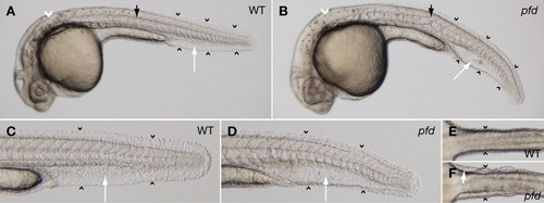
The pfdgw1 mutation disrupts notochord and vascular development. A: The notochord (black arrow), caudal vein (white arrow), and fin fold (black arrowheads) form normally in wild-type embryos, and melanin pigmentation is present (white arrowhead). B: pfdgw1 mutants exhibit notochord kinking (black arrow), a cavernous caudal vein with loss of the usual reticular venous plexus (white arrow), and fin fold attenuation (black arrowheads). Melanin pigmentation is present (white arrowhead). C: Fin fold (arrowheads) and caudal vein (arrow) in a wild-type embryo. D: Attenuated fin fold (arrowheads) and cavernous caudal vein (arrow) typical of pfdgw1 mutants. E,F: Ventral views of a wild-type embryo (E) and a pfdgw1 mutant (F) demonstrating skin distention secondary to edema in the mutant (F, arrowheads). Red blood cells have extravasated into the edematous area (F, arrow). All embryos were photographed at 30 hours postfertilization (hpf).
|