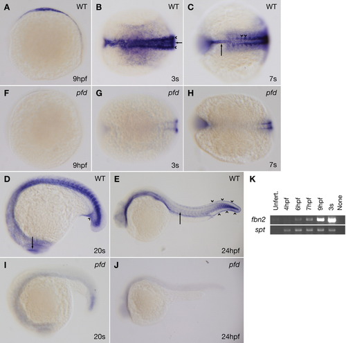
fbn2 expression is consistent with the pfdgw1 phenotype and dramatically reduced in pfdgw1 mutants. A-J: Clutches from pfdgw1/+ intercrosses were subjected to whole-mount in situ hybridization at the indicated developmental stages using probes to fbn2. A: Lateral view of a wild-type embryo at 9 hours postfertilization (hpf). B: Dorsal view of a wild-type embryo at the three-somite stage demonstrating fbn2 expression in the notochord (arrow) and paraxial mesoderm (arrowheads). C: Dorsal view of a wild-type embryo at the seven-somite stage demonstrating fbn2 expression in the notochord (arrow) and somites, with foci of increased staining near notochord-somite boundaries (arrowheads). D: Lateral view of a wild-type embryo at the 20-somite stage with fbn2 expression in the region of the developing caudal vein (arrowhead) and eye (arrow). E: Lateral view of a wild-type embryo at 24 hpf with hypochord (arrow) and prominent fin fold expression (arrowheads). F-J: fbn2 expression is dramatically reduced in pfdgw1 mutants at all stages analyzed. K: Reverse transcriptase-polymerase chain reaction (RT-PCR) for fibrillin-2 (fbn2) or spadetail (spt) using RNA from embryos at the indicated developmental stages. Unfert, unfertilized.
|
