|
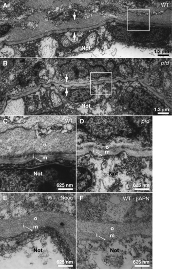
The outer layer of the notochord sheath is disrupted in pfdgw1 mutants. A-F: Transmission electron micrographs of truncal cross-sections from embryos at 30 hours postfertilization (hpf). A: Notochord sheath of a wild-type embryo (between arrows). The area in the white square is shown at higher magnification in panel C. B: Notochord sheath of a pfdgw1 mutant (between arrows). The area in the white square is shown at higher magnification in panel D. C: Notochord sheath of a wild-type embryo with inner (i), medial (m), and outer (o) layers. D: Notochord sheath of a pfdgw1 mutant where inner (i) and medial (m) layers are normal, but the outer (o) layer is reduced in size. E,F: Notochord sheaths of wild-type embryos treated with 10 μM neocuproine (E) or 10 mM &beta-aminopropionitrile (F). Not, notochord.
|