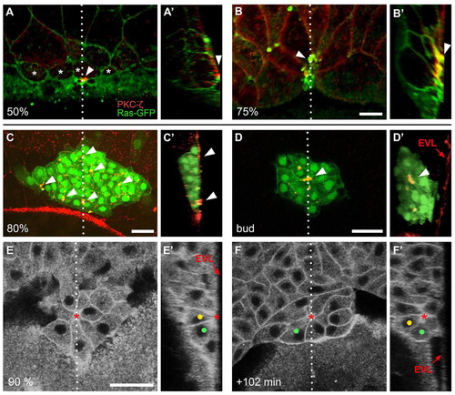
Apical attachment of the dorsal forerunner cell cluster to the enveloping layer and subsequent detachment to form a cellular rosette during gastrulation. (A-B′) Confocal images of Tg(β-actin:HRAS-EGFP) zebrafish embryos immunolabelled with an anti-aPKC-ζ antibody. GFP (green) and aPKC-ζ (red) channels are merged. Single dorsal focal planes (A,B) and sagittal sections at the position of the dotted line (A′,B′; embryo surface to the right) are shown. Animal pole is to the top. A surface plane through an embryo at 50% epiboly shows that presumptive DFCs (asterisks) are partially positioned underneath the EVL (A). Clear enrichment of aPKC-ζ is found at the contact point between one of the DFCs and the overlying EVL (A,A′; arrowheads). At 75% epiboly, the EVL completely covers the DFCs (B). A sagittal section shows bottle-shaped DFCs with strong aPKC-ζ enrichment at the contact points with the EVL (B′; arrowheads). (C-D′) Confocal images of Tg(sox17:GFP)-expressing embryos immunolabelled with an anti-ZO-1 antibody. GFP (green) and anti-ZO-1 (red) channels are merged. At 80% epiboly, DFCs form a compact cluster that is in contact with the interior surface of the EVL close to the margin. Anti-ZO-1 signal is enriched at contact points (arrowheads) between DFCs and the overlying EVL (dorsal z-projection is shown in C; sagittal section at the position of the dotted line in C′). At the bud stage, a ZO-1-rich focal point (arrowhead) is seen at the centre of the DFC cluster (single focal plane is shown in D; sagittal section at the position of the dotted line in D′). The cluster is disconnected from the overlying EVL. (E-F′) Images of a time-lapse multi-photon confocal movie of a Tg(β-actin:HRAS-EGFP) embryo, showing DFC cluster arrangement and tracing of two DFCs (green and yellow dots). Single dorsal focal planes (E,F) and sagittal sections at the position of the dotted line (E′,F′) are shown. At 90% epiboly, several bottle-shaped DFCs are attached to the overlying EVL via a single focal point (asterisk), including the two marked cells (E,E′; arrow indicates the position of the EVL). At +102 minutes, the marked cells have internalised and become part of a DFC rosette that is arranged around an interior focal point (F,F′; focal point marked by asterisk). Scale bars: 30 μm.
|
