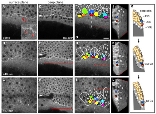
Dorsal forerunner cells are derived from dorsal surface epithelial cells. Images of a time-lapse multi-photon confocal movie of a zebrafish embryo expressing Tg(β-actin:HRAS-EGFP). Dorsal view with animal pole to the top. (A-C) Single focal planes through the surface of the embryo at early dome stage (∼4.2 hpf) and subsequent time points show the progression of dorsal surface epithelial (DSE) cells towards the vegetal pole. Some marginal DSE cells form filopodia-like protrusions at their leading edge (arrows and inset in A). (D-F) Images of the deep cell layer at equivalent time points to A-C, with the deep cell margin marked with arrowheads. DFCs (brackets) become visible directly in front of the deep cell margin shortly after the dome stage (E) and then move progressively away from the margin (F). (G-I) Fate mapping of DSE cells shows that nine of these cells at or close to the margin (coloured), but no deep cells, become converted into DFCs. Cells remaining within the enveloping layer (EVL) are outlined in white and traced by number. The same principal finding was made in all five embryos analysed by live imaging. The DSE-to-DFC conversion occurs as the DSE cells round up and become covered by the trailing EVL cells that form the new EVL margin. During this process, two of these cells (light green and dark blue) can be seen to divide once (H,I). During vegetal movement, both EVL cells and DFCs converge to the dorsal side. (J-L) Three-dimensional rendering and cutting of the image stacks along the dotted lines shown in G-I allows simultaneous display of the xy and yz axes, and reveals the repositioning of red and dark-blue DSE cells below the tissue. (M) Schematic representation of DSE-to-DFC conversion and deep cell internalisation as observed in the live recording. Two DSE cells, depicting the red and dark-blue DSE cells in the movie, become positioned beneath the EVL to become DFCs. The blue cell had divided immediately prior to internalisation. The attachment point linking DFCs to the EVL is shown as a yellow spot. Two deep cells (asterisks) are shown to undergo internalisation to form hypoblast. Scale bar: 30 μm.
|
