
Fig. 7
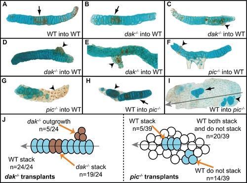
Cell autonomous behaviour of dak-/- and pic-/- chondrocytes. Transplanted dak-/- cells usually form columns with wild-type chondrocytes (arrow in B). However, in some cases dak-/- cells grow out from the wild-type host cells and behave autonomously (arrowheads in C,D,E). pic-/- cells transplanted into wild-type hosts never stack (arrowheads in F,G). In addition, wild-type cells both stack (arrows in H,I) and fail to stack (arrowheads in H,I) when transplanted into pic-/- hosts. Wild-type cells that stack in pic-/- hosts form columns that lie parallel to the longitudinal axis (grey arrow in I). (A–I) Dissected cartilage elements; all are ceratobrachial cartilage except (E) and (I) which are trabecular and ceratohyal cartilage respectively. Brown cells in (A–G) are transplanted cells, blue cells in (H,I) are transplanted cells. (J) summarises all 63 transplants analysed. Transplanted cells that flattened also intercalated to form columns. WT = wild-type. |