
Fig. 2
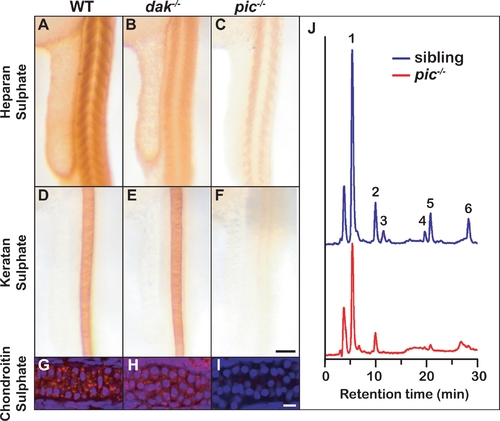
Sulphated proteoglycans are reduced in dak-/- and pic-/- larvae. Whole mount antibody staining at 24hpf reveals that HS is reduced in the somites of dak-/- and pic-/- (A–C) and KS in the notochord is reduced only in pic-/- larvae (D–F). Cartilage staining of the ceratohyal at 72hpf reveals that CS is made in wild-type and dak-/- chondrocytes (red stain in G,H), and absent from pic-/- cartilage (I) (nuclear staining is shown in blue with DAPI). HPLC analysis of HS in pic-/- larvae at day 5 indicates that sulphated disaccharides are nearly absent and unsulphated disaccharides are reduced (red) compared to their siblings (blue) (J). 1: ΔUA-GlcNAc, unsulfated Δ4,5-unsaturated hexuronate -N-acetyl glucosamine; 2: ΔUA-GlcNS, ΔUA-N-sulfated glucosamine; 3: ΔUA-GlcNAc6S, ΔUA-6-O-sulfated GlcNAc; 4: ΔUA-GlcNS6S, ΔUA-N-sulfated, 6-O-sulfated glucosamine; 5: ΔUA2S-GlcNS, 2-O-sulfated ΔUA-N-sulfated glucosamine; 6: ΔUA2S-GlcNS6S, 2-O-sulfated ΔUA-N-sulfated, 6-O-sulfated glucosamine. These disaccharides correspond to the major disaccharides found in both invertebrate and vertebrate animals. Panel F scale bar = 50μM. Panel I scale bar = 10μM. |
| Antibodies: | |
|---|---|
| Fish: | |
| Anatomical Terms: | |
| Stage Range: | Prim-5 to Protruding-mouth |