|
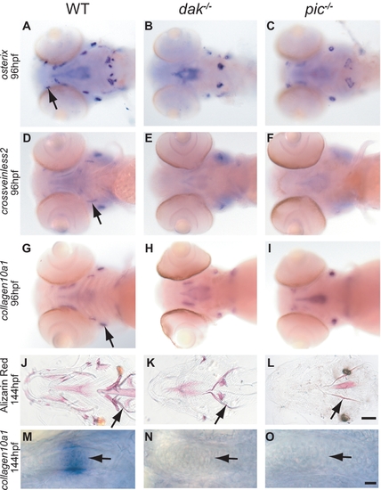
Reduction of bone development in dak-/- and pic-/- larvae. Wholemount RNA in situ analysis of osterix (A–C), crossveinless (D–F) and collagen10a1 (G–I) at 96hpf (all ventral views of the head). Markers for dermal and cartilage bone development are down-regulated or absent in both mutants (A,D,G wild-type; B,E,H dak-/-; C,F,I pic-/-). Arrows indicate wild-type expression in the maxilla (A), branchiostegal ray (D) and opercle (G). The loss of marker gene expression is consistent with the later reduction in bone formation in ventral views of 6 day old larvae stained with Alizarin Red (J,K,L). Arrows indicates the location of the cleithrum in J, K and L. Table S2 lists all of the affected bones. collagen10a1 expression in chondrocytes of the ceratohyal in wild-type larvae marks chondrocytes as they become hypertrophic (arrow in M). This expression is absent in dak-/- (N) and pic-/- (O) larvae. Panel L scale bar = 100μM. Panel O scale bar = 10μM.
|