
Fig. 4
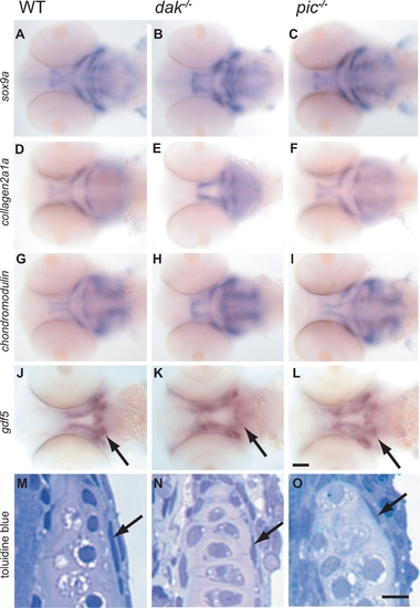
dak-/- and pic-/- larvae have wild-type levels of expression of markers of chondrocyte differentiation. Wholemount RNA in situ analysis of sox9a (A–C), collagen2a1a (D–F), chondromodulin (G–I) and gdf5 (J–L) at 60hpf (all ventral views of the head). Although the position of the developing skeleton varies between wild-type and mutants, the markers are expressed at similar levels in wild-type (A,D,G,J), dak-/- (B,E,H,K) and pic-/- (C,F,I,L). dak-/- larvae express sox9a at higher levels anteriorly, but this is perhaps due to more chondrogenic cells being present (see Figure 6). Expression of gdf5 in the perichondrium of the ceratohyal is present albeit slightly reduced in dak-/- and pic-/- (arrows in J–L). The perichondrium of the hyosymplectic is also seen in toluidine blue stained sections at day 5 (arrows in M,N,O). Panel I scale bar = 50μM. Panel L scale bar = 5μM. |
| Genes: | |
|---|---|
| Fish: | |
| Anatomical Terms: | |
| Stage: | Pec-fin |