
FIGURE
Fig. 5
Fig. 5
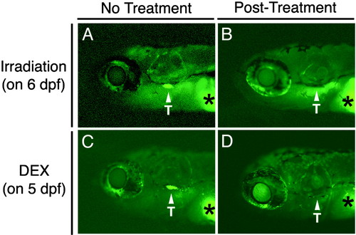
GFP-labeled T cells in 8-day-old lck-GFP fish are ablated in response to γ-irradiation or dexamethasone treatment. (A) Nonirradiated control fish. (B) Fish 2 days postirradiation. (C) Control fish with 0.4% ethanol. (D) Fish 3 days after treatment with 100 μg⋅ml-1 dexamethasone (DEX). Asterisks denote autofluorescence of the yolk sac. Arrowheads denote GFP-labeled cells in the thymus (T). The views are lateral with anterior to the left. |
Expression Data
Expression Detail
Antibody Labeling
Phenotype Data
Phenotype Detail
Acknowledgments
This image is the copyrighted work of the attributed author or publisher, and
ZFIN has permission only to display this image to its users.
Additional permissions should be obtained from the applicable author or publisher of the image.
Full text @ Proc. Natl. Acad. Sci. USA