
FIGURE
Fig. 3
Fig. 3
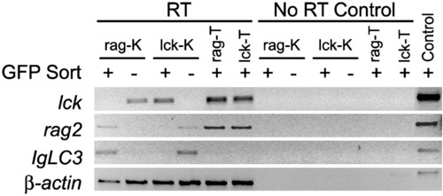
Semiquantitative RT-PCR analysis of FACS-sorted blood cell populations in the kidney (K) and thymus (T) of lck-GFP and rag2-GFP transgenic fish. GFP-positive (+) and -negative (-) blood cell populations are shown. Results for "No RT Control" show absence of genomic DNA contamination in samples. The β-actin PCR control was completed on genomic DNA. Because the β-actin primers span an intron, PCR amplifies a 100-bp larger fragment than seen in RT samples. |
Expression Data
Expression Detail
Antibody Labeling
Phenotype Data
Phenotype Detail
Acknowledgments
This image is the copyrighted work of the attributed author or publisher, and
ZFIN has permission only to display this image to its users.
Additional permissions should be obtained from the applicable author or publisher of the image.
Full text @ Proc. Natl. Acad. Sci. USA