
Fig. 5
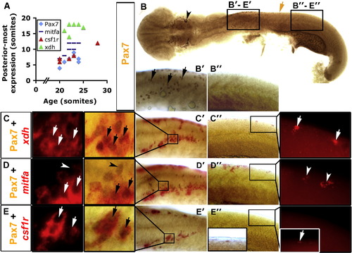
Xanthophore specification occurs before accumulation of Pax7. Immunodetection of Pax7 (brown, B–E) or in situ mRNA hybridisation (fast red fluorescence) for xdh (C), mitfa (D) or csf1r (E). Whole 20 s embryo flatmounts with anterior to left show dorsal and lateral views in head and trunk regions, respectively. Panels B′–E′ are anterior trunk, panels B″–E″ are posterior trunk according to position of black boxes in panel B. (A) Graph showing posteriormost extent of each marker in relation to embryonic stage. (B) Pax7+ NC are detected in the head (arrowhead) and in pre-migratory positions in the anterior trunk (black arrows in panel B′), but not in more posterior regions (B″). Brown arrow indicates the posteriormost extent of Pax7+ NC. (C–E) Dual stain for Pax7 and indicated mRNA showing anterior (C′, D′, E′) and posterior (C″, D″, E″) trunk in brightfield. The insets (C′–E′, C″–E″) are magnified to reveal fast red and its fluorescence. (C) xdh is expressed together with Pax7 (arrows) in anterior trunk, but alone in posterior trunk (C″ inset, white arrows). (D) mitfa is expressed in anterior and posterior trunk. Some Pax7+ NC co-express mitfa in anterior trunk, others do not (D′, arrows and arrowheads, respectively). mitfa is also expressed alone, probably in melanoblasts (Lister et al., 1999; D″, arrowheads). Mitfa expression is lateral to the Pax7+ cells in anterior trunk NC at this stage (D′). (E) csf1r mRNA is detected only in Pax7+ NC in anterior trunk (E′, arrows). However, the posteriormost Pax7+ NC are csf1r+ (E″, inset, white arrow). |
| Genes: | |
|---|---|
| Fish: | |
| Anatomical Terms: | |
| Stage: | 20-25 somites |
Reprinted from Developmental Biology, 317(2), Minchin, J.E., and Hughes, S.M., Sequential actions of Pax3 and Pax7 drive xanthophore development in zebrafish neural crest, 508-522, Copyright (2008) with permission from Elsevier. Full text @ Dev. Biol.