FIGURE

Fig. S3

ID
ZDB-FIG-080516-25
Publication
Shin et al., 2008 - Multiple roles for Med12 in vertebrate endoderm development
Other Figures
All Figure Page
Back to All Figure Page
Fig. S3

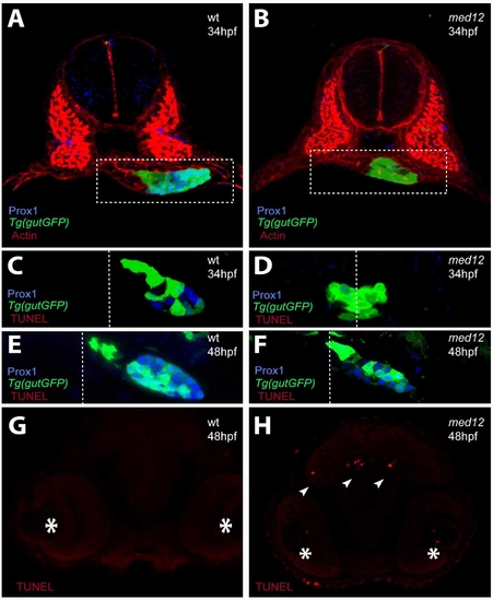
Analysis of apoptosis in med12s435 mutant embryos. (A, A′) Confocal projections of Prox1 (blue) and Actin (red) expression in wild-type and med12s435 mutants in the Tg(gutGFP)s854 line (green) at 34 hpf. White dashed lines surround the endodermal region. (C–F) Confocal projections of wild-type and med12s435 mutants in the Tg(gutGFP)s854 line (green) at 34 (C, D) and 48 (E, F) hpf in conjunction with anti-Prox1 (blue) antibody and TUNEL (red) staining. White dashed lines indicate the midline. In med12s435 mutants, Prox1 expression is mostly missing at 34 hpf but present at 48 hpf. Little if any TUNEL staining was observed in the endodermal tissues at these stages. (G–H) Confocal projections of TUNEL staining in the brain region of wild-type and med12s435 mutants. Asterisks indicate the eyes and arrowheads point to TUNEL-positive cells. Transverse sections, dorsal to the top.

Expression Data
Genes:
Antibody:
Fish:
Anatomical Term:
Stage Range: Prim-15 to Long-pec

Expression Detail
Antibody Labeling
Phenotype Data
Fish:
Observed In:
Stage Range: Prim-15 to Long-pec

Phenotype Detail
Acknowledgments
This image is the copyrighted work of the attributed author or publisher, and ZFIN has permission only to display this image to its users. Additional permissions should be obtained from the applicable author or publisher of the image.

Reprinted from Developmental Biology, 317(2), Shin, C.H., Chung, W.S., Hong, S.K., Ober, E.A., Verkade, H., Field, H.A., Huisken, J., and Stainier, D.Y., Multiple roles for Med12 in vertebrate endoderm development, 467-479, Copyright (2008) with permission from Elsevier. Full text @ Dev. Biol.