
Fig. 6
- ID
- ZDB-FIG-080514-23
- Publication
- Perz-Edwards et al., 2001 - Retinoic acid-mediated gene expression in transgenic reporter zebrafish
- Other Figures
- All Figure Page
- Back to All Figure Page
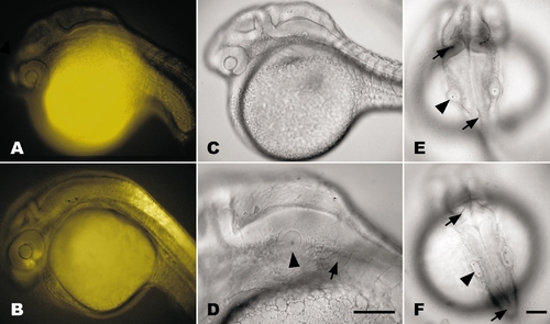
DEAB treatment at gastrulation abolishes transgene signal and teratogenizes zebrafish embryos by 24 hpf. (A) Treatment of transgenic embryo (RGYn2) with10 μM DEAB at shield stage (6 hpf) completely abolishes YFP expression by 24 hpf compared to DMSO-treated control (B). (C) DEAB treatment reduces frontonasal region, retina, hindbrain, and otic vesicle while inhibiting development of heart tube and fin bud. (D) Lateral view of the hindbrain demonstrates the significant reduction in the distance between the otic vesicle (arrowhead) and first myotome (arrow) in DEAB-treated embryo. (E and F) Dorsal views of DEAB-treated (E) and DMSO-treated (F) embryos demonstrate that the hindbrain is slightly reduced between the isthmus (top arrows) and the otic vesicles (arrowheads) while the caudal hindbrain between the otic vesicle and the termination of the ventricle (bottom arrows) is significantly reduced. The overall width of the hindbrain is also increased as a result of DEAB treatment. All embryos are oriented with rostral left and dorsal up, except for dorsal views in E and F, which have rostral up. Bars, 100 μm. Scale for A–C and E is the same as F. |
Reprinted from Developmental Biology, 229(1), Perz-Edwards, A., Hardison, N.L., and Linney, E., Retinoic acid-mediated gene expression in transgenic reporter zebrafish, 89-101, Copyright (2001) with permission from Elsevier. Full text @ Dev. Biol.