
Fig. 3
- ID
- ZDB-FIG-080514-20
- Publication
- Perz-Edwards et al., 2001 - Retinoic acid-mediated gene expression in transgenic reporter zebrafish
- Other Figures
- All Figure Page
- Back to All Figure Page
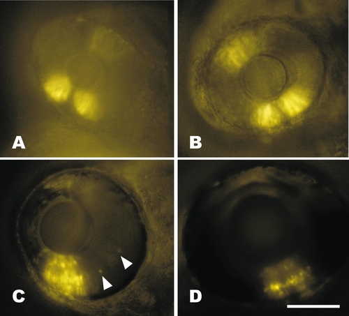
Time course of retinal expression in RGYn2. (A) Expression in the ventral retina patch is broader and stronger at 30 hpf than dorsal. Exposure time was 1 s. (B) Expression continues in similar dorsal and ventral patches by 48 hpf, but is much stronger. Exposure time was 0.25 s. (C) By 72 hpf the neural retina has differentiated into three distinct layers, all of which have transgene expression in RGY lines. A few expressing cells outside the ventral patch are visible in the inner nuclear layer (arrowheads). The dorsal retina is out of focus, but continues weak expression at this time. Exposure time was 0.1 s. (D) The layered expression continues in the ventral retina at 5 days postfertilization and occurs primarily in the inner and outer nuclear layers of the dorsal retina (out of focus). Exposure time was 0.5 s. Bar, 100 μm. All fish are oriented with rostral left and dorsal up. |
Reprinted from Developmental Biology, 229(1), Perz-Edwards, A., Hardison, N.L., and Linney, E., Retinoic acid-mediated gene expression in transgenic reporter zebrafish, 89-101, Copyright (2001) with permission from Elsevier. Full text @ Dev. Biol.