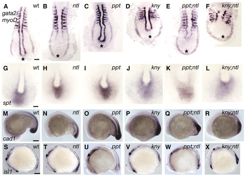
Posterior tissues are specified in ppt;ntl and kny;ntl mutants. At nine somites, myoD is expressed in the somites and adaxial cells, and gta2 is expressed in two bilateral stripes of prospective blood in wild-type (A) embryos. In ntl mutants (B), adaxial cell expression of myoD is lost and the stripes of gta2 are less separated posteriorly. In ppt mutants (C), adaxial cell expression of myoD is kinked and gta2 is similar to the wild type. In kny mutants (D), somites are mediolaterally broader and the AP lengths of adaxial cells and the gta2 domain are shorter. The ppt;ntl (E) and kny;ntl (F) mutants exhibit myoD expression patterns as expected for the combined individual mutants, and gta2 stripes are fused posteriorly. (G-L) Expression of spt in the paraxial mesoderm of wild-type (G), ntl (H), ppt (I), kny (J), ppt;ntl (K) and kny;ntl (L) embryos. (M-R) Expression of Caudal in the tailbud of wild-type (M), ntl (N), ppt (O), kny (P), ppt;ntl (Q) and kny;ntl (R) embryos. (S-X) Expression of isl1 in developing neurons at the 16 somite stage. Ectopic isl1 expression in observed in ntl (T), ppt;ntl (W) and kny;ntl (X) mutants. (A-L) Dorsal posterior flat mounts. (M-X) Lateral views. Scale bars=100 μm (A-F,M-X), 50 μm (G-L).
|
