
Fig. 4
- ID
- ZDB-FIG-080417-18
- Publication
- Blum et al., 2008 - Complex cell rearrangements during intersegmental vessel sprouting and vessel fusion in the zebrafish embryo
- Other Figures
- All Figure Page
- Back to All Figure Page
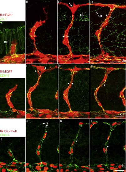
Dynamic expression of junctional proteins during ISV formation. Confocal projections of fli1:EGFP embryos labelled with anti-ZO-1 (A–D) or CDH5 (E–H) antibodies and of flk1:EGFPnls embryos labelled with CDH5 (I–L) antibody at different developmental stages. (A, E, I) 22–24 hpf, (B, F, J) 26–28 hpf, (C, G, K) 30 hpf, (D, H, L) 36 hpf. Already at early stages (A, E) both proteins are localized along the stalk of the ISVs, presumably between putative tip cells on the one hand, and between stalk cells on the other hand. Spots and lines (arrows in panels C, F and J) of ZO-1 and CDH5 are visible when tip cells start to extend in anterior and posterior direction to eventually form the DLAV. Arrowheads point to parallel junctions running along the axis of the ISV. Abbreviations: see Fig. 1, nt: neural tube. Scalebars: 20 μm. |
| Genes: | |
|---|---|
| Antibodies: | |
| Fish: | |
| Anatomical Terms: | |
| Stage Range: | 26+ somites to Prim-25 |
Reprinted from Developmental Biology, 316(2), Blum, Y., Belting, H.G., Ellertsdottir, E., Herwig, L., Lüders, F., and Affolter, M., Complex cell rearrangements during intersegmental vessel sprouting and vessel fusion in the zebrafish embryo, 312-322, Copyright (2008) with permission from Elsevier. Full text @ Dev. Biol.