
Fig. 2
- ID
- ZDB-FIG-080417-16
- Publication
- Blum et al., 2008 - Complex cell rearrangements during intersegmental vessel sprouting and vessel fusion in the zebrafish embryo
- Other Figures
- All Figure Page
- Back to All Figure Page
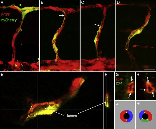
Endothelial cells are paired in the ISV. (A–D) Confocal projection of single cells expressing mCherry under control of the flk1 promoter (shown in green for better contrast) in fli1:EGFP transgenic embryos (red) in ISVs at 36 hpf (A–C) and 48 hpf (D) of development. (B) T-shaped cell embedded in the DLAV, (C, D) extended stalk cells. In all cases, mCherry-expressing cells appear to make up only part of the circumference of the tube (arrows), and appear paired or aligned with cells expressing EGFP only. (E) Single section (X–Y plane) of the cell shown in panel D. (F) Single cross section (Y–Z plane) of the ISV showing the lumen and the surrounding cells. (G, H) Single cross sections of ISVs at 48 hpf showing ZO-1 staining (arrows) between cells surrounding the lumen (2 cells in panel G, 3 cells in panel H). (G′, H′) Schematic representation of the cellular arrangement shown in panels G and H. Scalebars: 20 μm. |
| Gene: | |
|---|---|
| Fish: | |
| Anatomical Terms: | |
| Stage Range: | Prim-25 to Long-pec |
Reprinted from Developmental Biology, 316(2), Blum, Y., Belting, H.G., Ellertsdottir, E., Herwig, L., Lüders, F., and Affolter, M., Complex cell rearrangements during intersegmental vessel sprouting and vessel fusion in the zebrafish embryo, 312-322, Copyright (2008) with permission from Elsevier. Full text @ Dev. Biol.