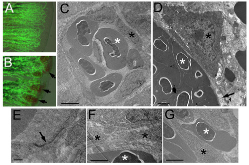
Cellular defects of reg6 endothelial cells. (A, B) Overlaid images of bright field and green fluorescent protein (GFP) of wild-type (A) and reg6 (B) 3-dpa regenerating fins showing the leakage of blood cells in reg6 fish (arrows). Transmission electron microscopic images of wild-type fish (C, low magnification; E, high magnification) showing that the blood vessel is enclosed by several endothelial cells which form nice, intact tight junctions (arrow in E). Additional images can be found in Additional file 1. In contrast, the blood vessels in reg6 fish (D) were usually leaking as evidenced by the lack of coverage by endothelial cells (arrow) and the presence of blood cells outside blood vessels (as an endothelial cell is sandwiched by two blood cells in G and arrows in Additional file 2). In addition, reg6 endothelial cells appeared less stretched-out (D), and tight junction were rarely formed among them (F). Scale bars, 2 μm for C, D, F, and G; 10 nm for E. white asterisks, blood cell; black asterisks, endothelial cell.
|
