
Fig. 1
- ID
- ZDB-FIG-080326-105
- Publication
- Devine et al., 2008 - Robo-Slit interactions regulate longitudinal axon pathfinding in the embryonic vertebrate brain
- Other Figures
- All Figure Page
- Back to All Figure Page
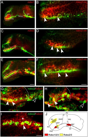
Robo family members are expressed in the brain during the formation of the axon scaffold. Lateral views of whole mount zebrafish brains double labeled for the expression of robo family members (red) and the HNK-1 epitope (green) at 22 h post-fertilization (hpf). Rostral is to the left and dorsal is to the top. (A) At 22 hpf, robo1 expression is observed in the ventral diencephalon (dashed outline), ventrocaudal cluster (vcc) and midbrain. (C) robo2 expression was observed in the ventrorostral cluster (vrc; dashed outline), dorsorostral cluster (drc) and vcc. (E) robo3 expression was detected in the vrc (dashed outline), drc and vcc. Single slice confocal analysis of robo expression reveals co-localization of individual robo receptors to vrc cell bodies at 22 hpf (B, D and F, filled arrowheads). (G–I) Double in situ hybridization for robo family members in the rostral diencephalon at 22 hpf, rostral is to the left, dorsal is to the top. robo1 co-localizes with robo2 (G, filled arrowheads) and robo3 (H, filled arrowhead) in a subset of rostrally positioned cells (dashed outline defines the caudal limit of co-expression). Outside of this region of overlap, additional cells were identified that expressed only one of the robo receptors (G and H, unfilled arrowheads). robo3 co-localizes with the majority of robo2-expressing cells (I, filled arrowhead) and is also detected in surrounding cells (I, asterisk). (J) Schematic representation of robo1, -2 and -3 mRNA expression at 22 hpf. Scale bars: A, C, E: 50 μm; B, D, F: 10 μm; G–I: 12.5 μm. |
| Genes: | |
|---|---|
| Fish: | |
| Anatomical Terms: | |
| Stage: | 26+ somites |
Reprinted from Developmental Biology, 313(1), Devine, C.A., and Key, B., Robo-Slit interactions regulate longitudinal axon pathfinding in the embryonic vertebrate brain, 371-383, Copyright (2008) with permission from Elsevier. Full text @ Dev. Biol.