
Fig. 2
- ID
- ZDB-FIG-071112-3
- Publication
- Kosaka et al., 2007 - Spatiotemporal localization of germ plasm RNAs during zebrafish oogenesis
- Other Figures
- All Figure Page
- Back to All Figure Page
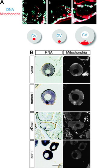
Co-localization of germ plasm RNAs with the mitochondrial cloud in zebrafish oocytes. (A) Stage I oocyes were stained with Mitotracker Red580 (mitochondria) and DAPI (DNA). Schematic representation was shown at the bottom of each panel. In panels a and b, a mitochondria cloud was found adjacent to the germinal vesicle (GV), whereas mitochondria staining was observed at or near the vegetal cortex in panel c. (B) The right panel shows mitochondria staining of a section of a stage I oocyte. For the left panel, in situ hybridization was carried out on the same section as that shown in the right panel using the vasa, nanos1, zDazl and zorba/ZOR-1 probes. Sections were cut at a thickness of 10 μm. Scale bars, 50 μm. |
| Genes: | |
|---|---|
| Fish: | |
| Anatomical Term: | |
| Stage: | Adult |
Reprinted from Mechanisms of Development, 124(4), Kosaka, K., Kawakami, K., Sakamoto, H., and Inoue, K., Spatiotemporal localization of germ plasm RNAs during zebrafish oogenesis, 279-289, Copyright (2007) with permission from Elsevier. Full text @ Mech. Dev.