
Fig. 5
- ID
- ZDB-FIG-071112-6
- Publication
- Kosaka et al., 2007 - Spatiotemporal localization of germ plasm RNAs during zebrafish oogenesis
- Other Figures
- All Figure Page
- Back to All Figure Page
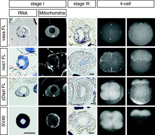
The 3′ UTRs of vasa, nanos1 and dazl RNAs govern localization during oogenesis and early embryogenesis. In situ hybridization of GFP mRNA fused with vasa, nanos1, dazl, or SV40 3′ UTRs expressed in transgenic zebrafish oocytes (stages I and III) and fluorescein in situ hybridization in 4-cell embryos. GFP-nanos1 mRNA was expressed by zpc promoter (Onichtchouk et al., 2003), whereas other mRNAs were expressed by EF1α promoter. In stage I oocytes, in situ hybridization (RNA) was performed on sections counter-stained with Mitotracker (Mitochondria). Oocyte sections were cut at a thickness of 10 μm. Scale bars, 50 μm. Lateral and animal views of the 4-cell embryo are shown. |
Reprinted from Mechanisms of Development, 124(4), Kosaka, K., Kawakami, K., Sakamoto, H., and Inoue, K., Spatiotemporal localization of germ plasm RNAs during zebrafish oogenesis, 279-289, Copyright (2007) with permission from Elsevier. Full text @ Mech. Dev.